Pregnancy
Everything you need to know
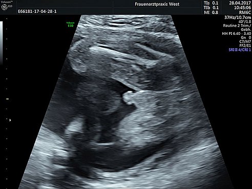
Combined First Trimester Screening
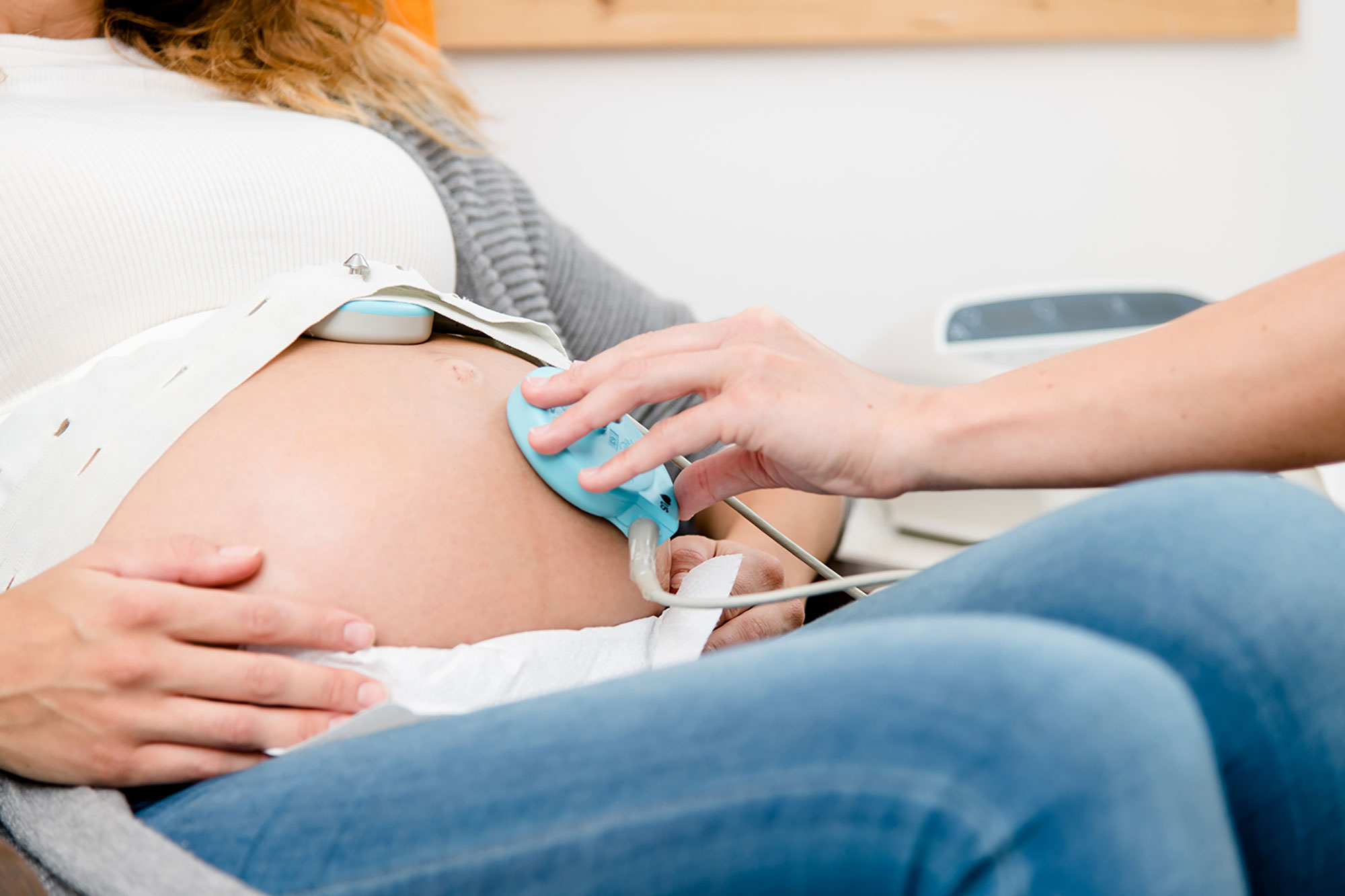
The first trimester screening (nuchal translucency screening), one of the most important ultrasound scan during pregnancy is performed between 11 and 13 weeks of pregnancy.
In the first part of the scan we calculate the risk of the fetus having extra chromosomes, called trisomies. This test uses a combination of the mother´s age, hormones that come from the placenta and a sonogram measuring the pocket of fluid at baby´s neck. The screening test has a 90-93 % detection rate of Down-syndrome (Trisomy 21) and a 5 % false positive rate.
In the second part follows the evaluation of the whole fetus´s anatomy as far as possible in this early week of gestation. Most of the major anomalies can be already detected at this stage, but the main goal of the scan is to confirm normal development and reassure parents.
Thirdly the screening can be completed with a risk estimation for developing a preeclamspia after 20 weeks.
The screening can only be performed by physicians who are yearly certified. Dr. Jánky is certified for First Trimester Screening through the Fetal Medicine Foundation (FMF) in London.
Cell-Free Fetal DNA Screening (NIPT)
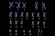
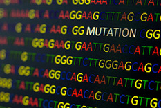
With a DNA-based blood screening test you can detect chromosomal anomalies, as well as detect the gender. The test can exlude following conditions: Trisomies 13, 18 and 21, Turner-Syndrome, Klinefelter-Syndrome, Triple-X-Syndrome and XYY-Syndrome.
The test can be performed as early as 9th week of pregnancy. It has a very high detection rate for chromosomal anomalies (99,8 % for Down-Syndrome) but gives no hint at all about structural malformations of the fetus.
Thus, according to current recommendations, the NIPT is usually performed together with the first trimester screening.
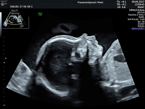
Genetical Ultrasound
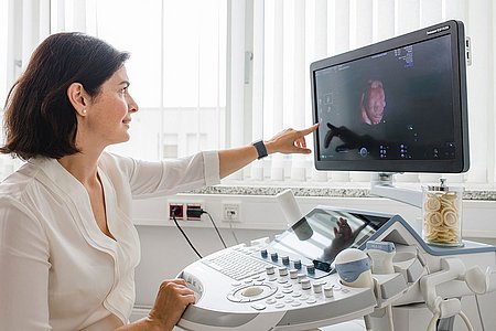
At this scan between 20 and 22 weeks of pregnancy we check baby´s anatomy again, ideally as the second „full“ ultrasound scan after the first trimester screening. All the organs of the fetus are examined systematically, from brain and face down to the heart and extremities.
Furthermore we check the amount of amniotic fluid, the placenta as well as the blood flow in some arteries.
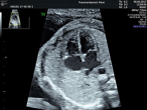
Fetal Heart Ultrasound Scan
With a heart scan we check the baby´s heart in detail with a high resolution ultrasound maschine. In the vast majority of cases we rule out a heart condition. However in a few cases we diagnose a heart failure. The accurate diagnose allows an early consultation with pediatric cardiologists and pediatric heart surgeons, to provide optimum care before and after birth. In most of these cases we plan the delivery in a pediatric heart center with appropriate specialists.
When is the scan performed?
In principle, fetal heart can be checked beyond 12-13 weeks of pregnancy. About 50-60 % of heart conditions can be then detected. However the reliability of the scan at this early stage of pregnancy is limited due to small size of the heart, so that we recommend another scan later in pregnancy. The most favourable time for the heart scan is between 20-22 weeks, where specialists can detect up to 80 % of all heart conditions.
Of course a fetal heart scan can be performed at any stage of pregnancy by suspicion of a heart failure, cardiac arrhythmia or in existence of other malformations.
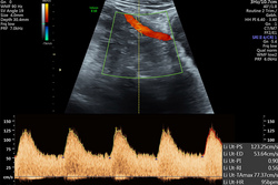
Blood Flow Measurement
The Doppler scan is a special ultrasound technique to measure blood flow velocitiy and resistance in the vessels of the uterus as well as in baby´s vessels and organs. From the obtained data conclusions can be drawn about the adequate supply of the child with nutrients and oxygen, as well as on cardiac and circulatory function.
Most commonly we measure the blood flow in the umbilical cord and brain arteries of the baby and in the uterine arteries of the mother.
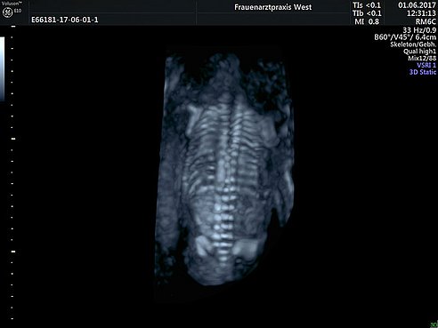
3D/4D Fetal Imaging
The 3D ultrasound is an ultrasound examination which is extended by one dimension opposite the two-dimensional and thus it is possible to represent the unborn child as well as its body parts spatially. In the case of the 4D ultrasound, the time is added as a 4th dimension, ie the three-dimensional image is generated in real time. For this reason, the 4D ultrasound is also called live 3D ultrasound. This results in a constantly updated, three-dimensional image on the monitor in which the child movements can be displayed in real time.
The examination can be used as a supplement to the conventional ultrasound examination for special questions, to the exclusion or detection of some malformations, but of course also at the request of the pregnant woman.
When is the best time for the scan?
The most beautiful pictures can be made in the second trimester between 24.-30. pregnancy week. The quality of the images depends on various factors, e.g. the amount of amniotic fluid, the thickness of the maternal abdominal wall, the location of the placenta and the baby´s position.
What should you pay attention to?
Please do not put any creams or lotions on your belly on the day of the examination.
How long does the scan take?
The 3D / 4D ultrasound examination takes 15-45 minutes depending on the ultrasound conditions and indication.
Make an appointment