
Breast cancer detection in women with dense breast tissue using
3D breast ultrasound.
Breast cancer detection in women with dense breast tissue using
3D breast ultrasound.
With the Invenia™ ABUS Premium, we offer a modern form of automated 3D breast ultrasound. We recommend this examination to women aged 30 and older as a valuable addition to breast cancer screening. ABUS is particularly beneficial for women with dense breast tissue, as it aids in early detection and effectively complements mammography.
During the procedure, the entire breast tissue is captured in a standardized, three-dimensional manner—without radiation exposure. This allows for precise documentation of any findings and reliable comparison during future examinations.
Of course we still offer conventional handheld breast ultrasound (2D) in our clinic. However, due to limited capacity for this method, we provide 3D breast ultrasound (ABUS) as a modern alternative that ensures a standardized examination of the entire breast. Many patients opt for ABUS because it delivers exceptional image quality and enables a reliable assessment of the entire breast tissue.
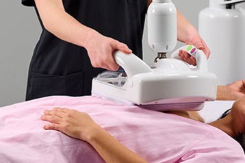
The Invenia™ ABUS Premium (Automated Breast Ultrasound System, ABUS) is a state-of-the-art system for automated 3D breast ultrasound. Unlike conventional handheld ultrasound, the entire breast is scanned in a standardized, automated manner.
A specialized scanner captures high-resolution, three-dimensional images of the entire breast tissue. All images are subsequently personally evaluated by Dr. Jánky. Thanks to the standardized imaging process, the scans can be easily compared with those from future examinations.
The procedure involves no radiation, causes minimal discomfort, and takes only a few minutes. It serves as a valuable addition to breast cancer screening—particularly for women with dense breast tissue. Evaluations are performed exclusively by Dr. Jánky, who holds DEGUM Level II certification in breast ultrasound.
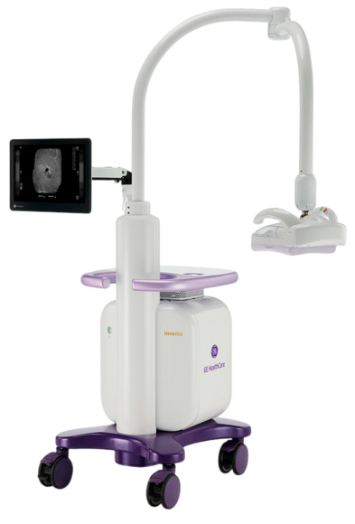
| Conventional Breast Ultrasound | Invenia™ ABUS Premium |
| Manual examination | Automated examination |
| Individual image sections | 3D view of the entire breast |
| Examiner-dependent | Standardized imaging |
| Limited reproducibility | Comparable follow-up assessments |
| 2D imaging | Modern 3D technology |
3D breast ultrasound (ABUS) is a modern, radiation-free supplement to breast cancer screening. Standardized three-dimensional imaging can assist in the assessment of breast tissue—particularly in cases of dense breast tissue—and provide additional diagnostic information.
The entire breast is captured and displayed in three dimensions. This allows for reliable comparison of changes during follow-up examinations. The procedure is gentle, takes only a few minutes, and does not require the intense compression associated with mammography.
Imaging without radiation exposure
3D imaging of the entire breast
Standardized and reproducible imaging
Good comparability for follow-up assessments
Particularly beneficial for dense breast tissue
Gentle examination without strong compression
Valuable complement to mammography
We recommend 3D breast ultrasound (ABUS) once a year for women aged 30 and older as a supplement to the manual examination during gynecological cancer screening. The examination can aid in early detection, particularly in cases of dense breast tissue.
ABUS can also be a useful option:
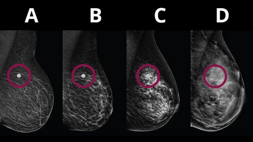
Approximately 40% of women have dense breast tissue. Since both dense breast tissue and tumors appear bright on a mammogram, abnormalities can be more difficult to detect. In these cases, 3D breast ultrasound can usefully complement mammography.
We will discuss which examinations are recommended for your specific situation during a personal consultation.
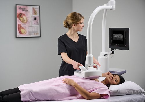
The examination is performed while you lie comfortably on your back and takes only a few minutes per breast. First, a specially trained staff member applies ultrasound gel to the breast. Next, the breast is gently scanned using the ABUS scanner, which automatically captures a three-dimensional image. The second breast is then examined in the same way.
The procedure is completely radiation-free and painless. No strong breast compression is required—only light pressure from the scanner, which most patients find comfortable.
All images are subsequently carefully evaluated personally by Dr. Jánky (DEGUM Level II). You will receive a written report. Should any findings appear abnormal or inconclusive, we will promptly invite you for a follow-up examination using conventional handheld breast ultrasound and a personal consultation with the doctor. Any further steps will then be discussed with you individually.
No. Automated 3D breast ultrasound (ABUS) is a supplementary ultrasound examination of the breast and does not replace mammography.
Mammography remains a key component of early detection—particularly within the framework of statutory breast cancer screening programs for women aged 50 and older.
For younger women and those with dense breast tissue, an additional ultrasound examination may be beneficial. In our practice, we offer both conventional (2D) breast ultrasound and automated 3D breast ultrasound (ABUS) for this purpose; the latter enables a highly standardized and comprehensive view of the breast tissue.
Yes. The examination is performed entirely without X-rays.
The examination is generally perceived as gentle and well-tolerated.
Dense breast tissue is very common and is not a sign of disease. It simply describes the composition of the tissue in the breast.
When breast tissue is dense, it can sometimes be more difficult to make a clear assessment during a mammogram. This is because dense tissue and potential abnormalities can look similar.
For this reason, an additional ultrasound scan may be useful. 3D breast ultrasound (ABUS) shows the breast tissue from a different perspective and can provide additional information for the assessment.
Approximately 40% of women have dense breast tissue.
An examination costs €150 for patients with statutory health insurance. Billing for self-payers and privately insured patients is based on the GOÄ (Official Scale of Medical Fees).
You should allow for a total of about 30 minutes for the examination.
The actual image acquisition for each breast takes only a few minutes. This is in addition to the time required for applying the ultrasound gel, positioning, and switching between the two sides.
You may leave the clinic immediately after the examination. Dr. Jánky will subsequently evaluate the images, and the written report will be made available to you. Should any abnormal findings arise, you will, of course, be promptly scheduled for a follow-up consultation or additional examination.
If cancer is detected at this early stage, chemotherapy can be avoided in 70% of patients.1
The examination using the Invenia™ ABUS Premium costs €150 for patients with statutory health insurance.
For privately insured patients and self-payers, billing is based on the official scale of fees for physicians (GOÄ).
Please note: 3D breast ultrasound (ABUS) is classified as an individual health service (IGeL) and is not currently covered by statutory health insurance plans. Consequently, the costs are generally not reimbursed by statutory health insurance providers.
Please feel free to contact us if you have any questions regarding costs or billing. We provide transparent advice and will, of course, inform you of the applicable costs prior to the examination.

Would you like a consultation regarding automated 3D breast ultrasound or wish to schedule an appointment?
The Frauenarztpraxis West in Regensburg would be happy to provide you with personal information about modern breast diagnostic options using Invenia™ ABUS Premium.
Simply get in touch with us — we are here to help!
Tel: 0941 41171